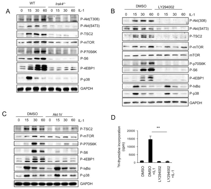
Figure 1. PI3K and AKT are required for IL-1-induced mTOR activation.
(A) Cell lysates from wild-type and Irak4−/− Th17 cells untreated or treated with IL-1 (10ng/ml) for different time points were analyzed by protein blot analysis using antibodies as indicated. (B–C) Wild-type Th17 cells were untreated or treated with IL-1 (10ng/ml) for different time points in the presence and absence of (B) 10μM PI3K inhibitor (LY294002) and 1μM Akt inhibitor (Akt IV) (C). Cell lysates were analyzed by protein blot analysis using antibodies as indicated. (D) Wild-type Th17 cells were rested for overnight, followed by incubation with 10ng/ml IL-1 in the presence and absence of 10μM PI3K inhibitor (LY294002). The treated cells were incubated one additional day with 3H for thymidine incorporation experiment. Error bars, s.d.; **, p<0.01 (two tailed t-test). Data are representative of three independent experiments.

