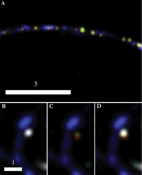
Fig. 2.
BDNF-mCherry and tPA-EYFP are copackaged in presynaptic DCGs. Deblurred fluorescence image (A) of an axon from a 14-DIV hippocampal neuron showing colocalization of tPA-EYFP (green) and BDNF-mCherry (red) within DCGs that, in turn, colocalize with SV clusters (blue) immunostained against synapsin-1 using a Cascade Blue secondary. DCGs containing tPA and BDNF appear yellow. Images (B-D) of a synapse between fixed 21-DIV hippocampal neurons. DCG proteins are tagged as in (A) and the SV cluster (white) again is immunostained against synapsin-1, but with a secondary, Alexa Fluor 647, that emits in the far red. The spine (blue) is labeled using an EBFP2 fill. For clarity, the spine is shown first with the SV cluster (B) and then with the two overlapping DCG proteins (C). All stained components are shown together in (D). Scale Bar = 5 μm for A. Scale Bar = 1 μm for B-D.

