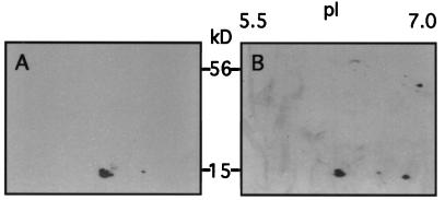
Figure 5.
Antibody detection of S from enriched Rubisco samples resolved in 2-D gels. S-specific antibodies were used to probe protein blots of 2-D gels. Chemiluminescence was used to produce fluorographs displaying S positions. A, Antibody detection of S in wild-type Arabidopsis treated with blue light. Three S spots are apparent. B, Antibody detection of S in T7.5 under white light. A fourth S is resolved. Also shown in B is an unidentified spot at 45 kD with a pI of 7, which is neither S nor its precursor.

