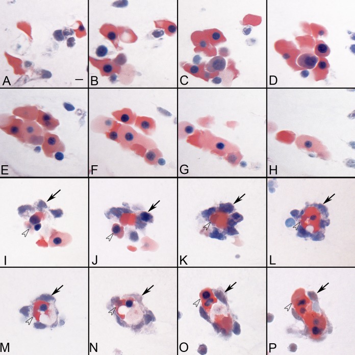
Figure 2 .
Serial sections showing two types of BI-like structures in the vitreous at 5.5 WG. Some of the BIs consisted of aggregates of erythroblasts (A–H), while others (I–P) were composed of basophilic mesenchyme (arrows) surrounding a core of highly acidophilic erythroblasts (arrowheads). Wrights-Giemsa stain, Scale bar in A = 10 μm for all.

