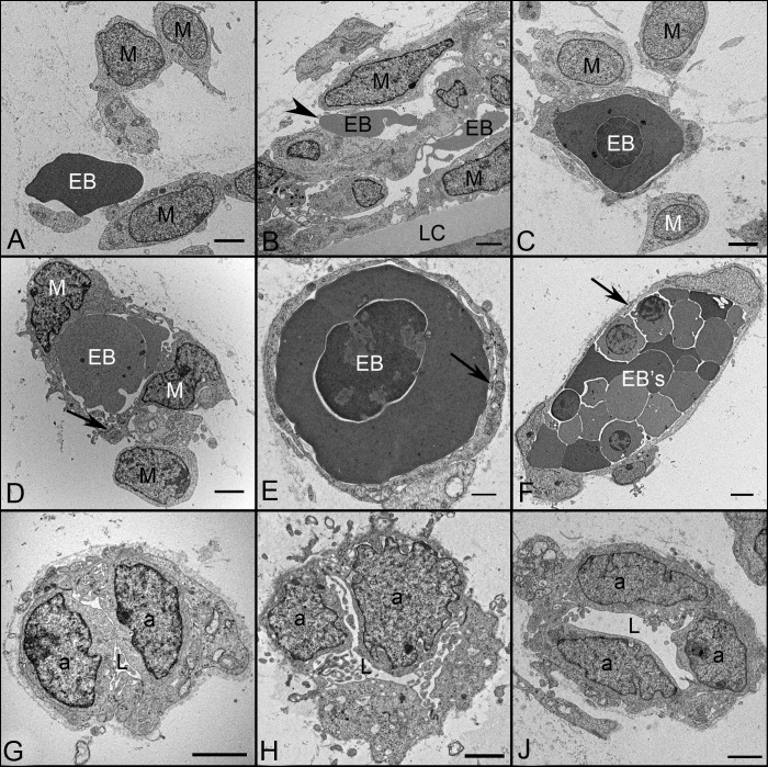
Figure 3. .
Ultrastructural features of BIs (A–F) and angioblastic aggregates (G–I) in the vitreous of a 6.5 WG embryonic eye showing mesenchymal cells (M), erythroblasts (EB), angioblasts (A), and early lumen formation (L). BIs (A–C) were composed of loosely arranged mesenchymal cells with free erythroblasts (A), incompletely enveloped erythroblasts (B), and erythroblasts completely enveloped by mesenchymal cells (C). In more mature structures (D–F), the central core of erythroblasts were completely surrounded by mesenchymal cells connected by junctions (arrows). Angioblastic aggregates (G–H) initially formed cords with slit-like lumen and finally primordial capillaries with lumen (J). There was a paucity of basement membrane material surrounding these BI and primitive capillaries. Scale bars = 2 μm in all.

