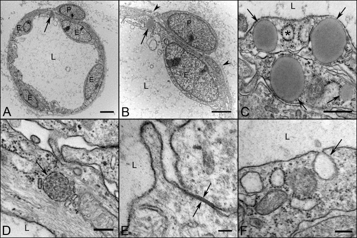
Figure 4. .
Ultrastructural appearance of capillaries and their cellular components at 12 WG. Capillaries (A, B) have well differentiated endothelium (E) and pericytes (P). The number of endothelial cells profile per capillary far outnumbered the perictytes. The lumen were fully open (L in all) and there was a continuous thin basement membrane present (arrowheads in B). Endothelial cells (C–F) contained lipid droplets (arrows in A–C), and coated pits (asterisk in C). Weibel-Palade bodies (arrow in D) were frequently observed at this age, as were tight junctions (opposing arrows in E) and caveolae (arrow in F). Scale bars = 2 μm in A, B, 500 nm in C, 250 nm in D, F, and 100 nm in E.

