Figure 6. .
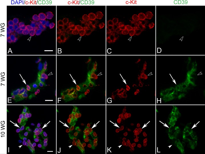
Double-labeling for c-Kit and CD39 in BIs of vitreous at 7 WG (A–H) and blood vessels at 10 WG (I–L). At 7 WG, free erythroblasts and aggregates (arrowhead in A–D) expressed c-kit in their cytoplasm or membrane, but did not express CD39. All mesenchymal cells in BI were double-labeled with c-Kit/CD39 and some select cells showed nuclear c-Kit immunoreactivity (arrow in E–H). Free erythroblasts outside BI were CD39− (arrowhead in E–H). At 10 WG, all cells of blood vessels were c-Kit/CD39+ (I–L) with c-Kit being expressed exclusively in nuclei (arrows). A similar pattern of staining was observed at 12 WG (not shown). Scale bars in A, E, I = 10 μm.

