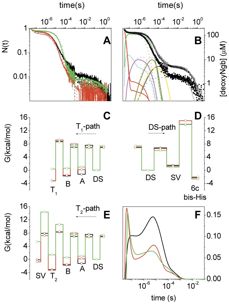
Figure 3. Laser-flash photolysis of D. mawNgb*, C. aceNgb* and human Ngb.
A. Comparison between CO-rebinding kinetics measured at 436 nm for D. mawNgb* (red), C. aceNgb* (black) and human Ngb (green) at 20°C (solid lines) and 10°C (dotted lines). Solutions were equilibrated with 1 atm CO. B. Fitting of CO-rebinding curves to C. aceNgb* T = 20°C, 1 atm (filled circles), 0.1 atm (open circles). Reaction intermediates are also reported as solid and dotted lines for data taken at 1 atm CO and 0.1 atm CO, respectively. C, D, E. Free-energy profiles at 20°C for ligand migration through the internal cavities, ligand exit to the solvent from the distal pocket, and six-coordination by distal His. In black, C. aceNgb*; in red, D. mawNgb*; in green, human Ngb. F. Time course of the fraction of photodissociated ligands migrating through cavities as estimated from the fitting with Scheme 1. In black, C. aceNgb*; in red, D. mawNgb*; in green, human Ngb. T = 20°C, 1 atm CO.

