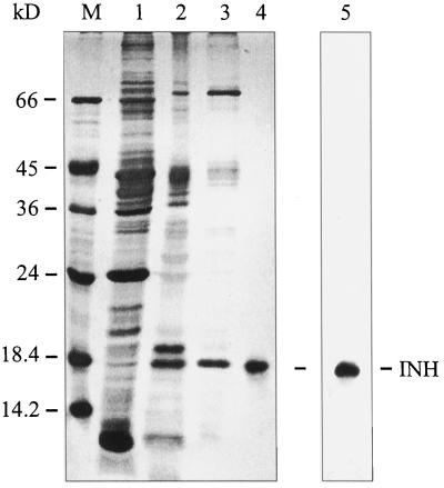
Figure 1.
Purification of tobacco INH from a salt-eluted cell wall protein fraction from transformed tobacco cells. Lane M, Marker proteins; lane 1, ammonium sulfate fraction (40–85% saturation); lanes 2 and 3, CWI activity peak fractions from cation-exchange chromatography, pH gradient (lane 2) and NaCl gradient (lane 3; Weil and Rausch, 1994); lane 4, electroeluted INH protein from the NaCl-gradient peak fraction (see lane 3); lane 5, immunoblot of ammonium sulfate fraction (see lane 1) with affinity-purified polyclonal antiserum directed against INH.

