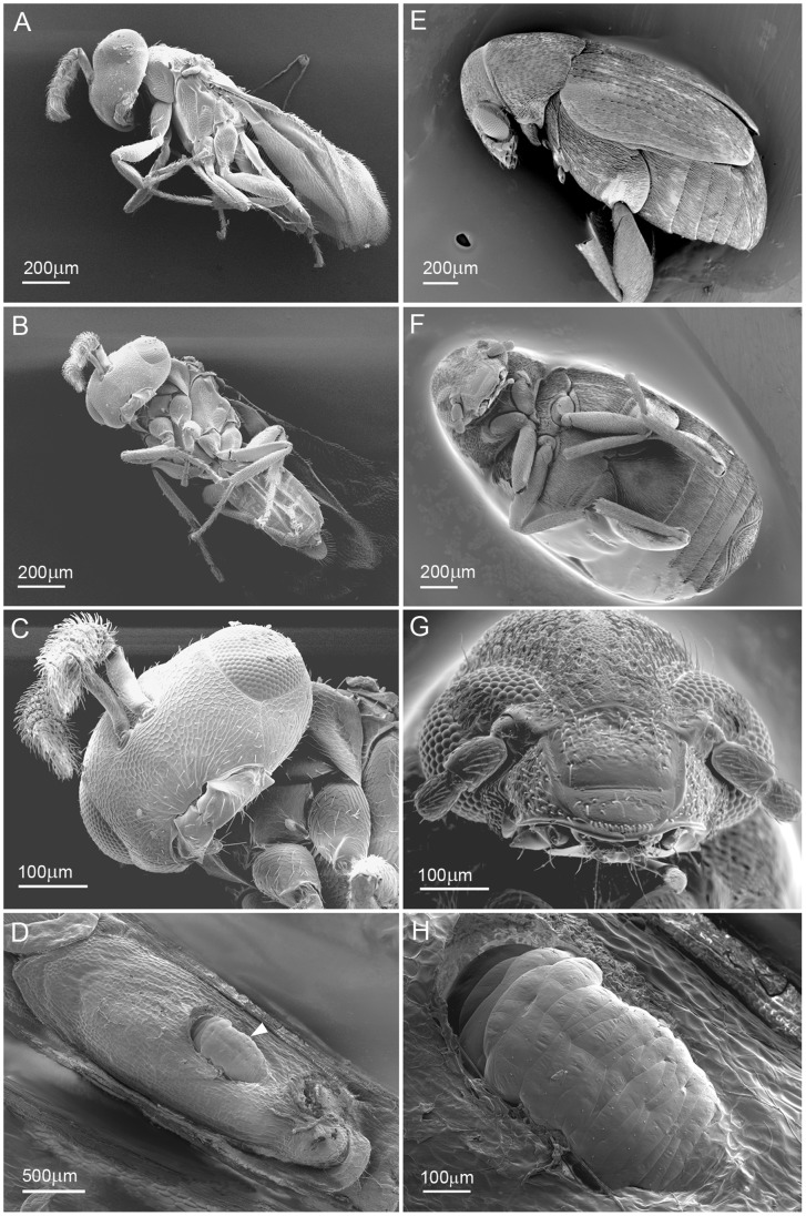
Figure 3. Scanning Electron Micrographs of seed chalcid wasp, seed chalcid wasp larva (in situ Stanleya pinnata seed) and seed beetle.
Images A–C are views of the chalcid wasp, areas around the mouth, thorax, legs and abdomen were targeted for Se using EDS. On these external surfaces only very low levels of Se were detected from upper leg segment spines (more detail is provided in Material S1). The seed chalcid wasp larva in image D (white arrow head) & H at higher magnification gave low positive Se signals around a spiracle and also on some bristles/spines seen on the external surface (Material S1). Images E – G are views of the seed beetle, which was also targeted for Se around the mouth, legs and abdomen using EDS, with no positive signals detected.

