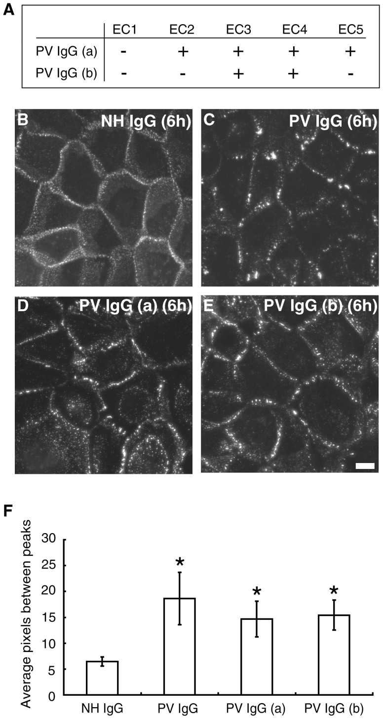
Figure 7. PV IgG lacking antibodies directed against the Dsg3 EC1 domain cause Dsg3 clustering.
(A) Two PV patient samples, PV IgGa (patient #3270) and PV IgGb (patient #7860) were identified in which epitope mapping revealed the absence of antibodies directed against the N-terminal extracellular domain (EC1) of Dsg3. (B–E) Cell surface Dsg3 was monitored using biotinylated AK23. NH IgG caused no change in the distribution of cell surface Dsg3 (B) whereas a typical PV IgG containing EC1 domain antibodies caused Dsg3 clustering (C). Both PV IgG(a) and PV IgG(b) caused Dsg3 clustering (D,E). (F) Quantitative assessment of clustering. *Indicates statistical significance compared to NH IgG (P<0.05). Scale bar, 10 µm.

