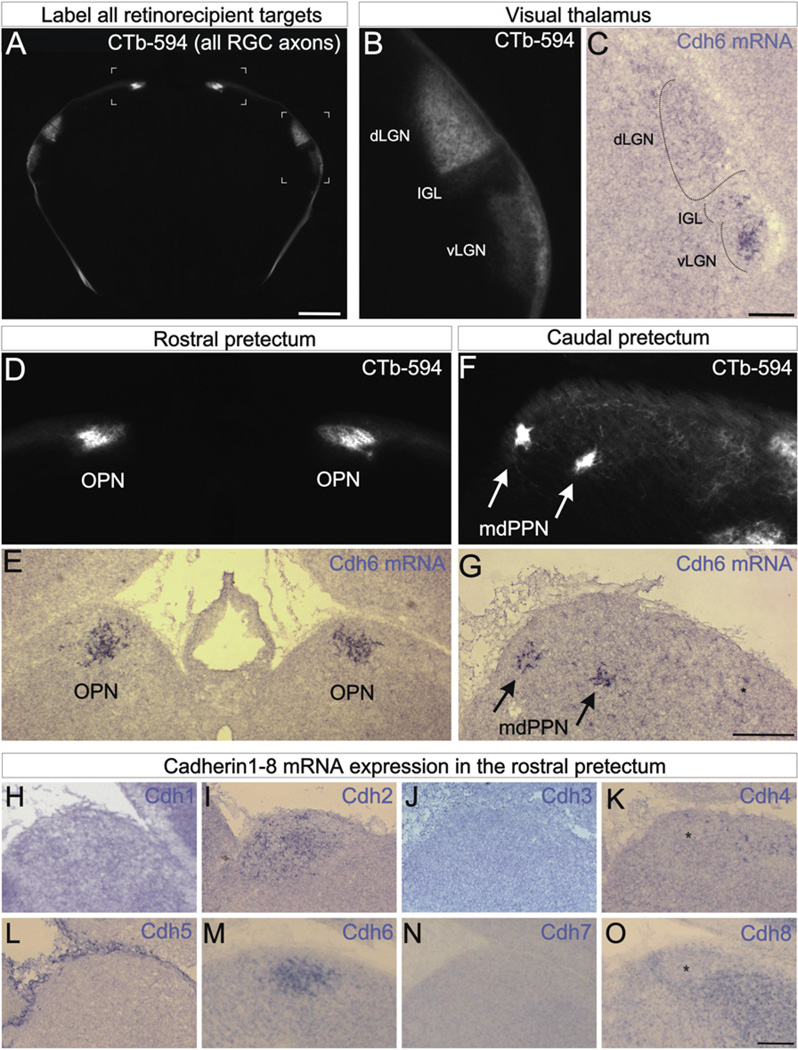
Figure 1. Cadherin-6 Is Expressed in Specific Subcortical Visual Nuclei.
(A) CTb-594 labeled RGC axons at the forebrainmidbrainborder of a postnatal day 2 (P2) mouse. Image is in coronal plane. Bracketed regions correspond to panels (B and D). Scale = 500 µm. (B) Pan-RGC axon labeling in the visual thalamus. The ventral lateral geniculate nucleus (vLGN), intergeniculate leaflet (IGL) and dorsal lateral geniculate nucleus (dLGN) contain RGC axons. (C) Cdh6 mRNA expressing cells in the IGL and vLGN. Scale = 100 µm. (D) CTb-594 labeled RGC axons in the rostral pretectum. The left and right olivary pretectal nuclei (OPN) are densely innervated. (E) Cdh6 mRNA expression in the OPN of a P1 mouse. (F) RGC axons in the caudal pretectum. The medial division of the posterior pretectal nucleus (mdPPN) of Scalia (1972) appear as two foci (arrows). (G) Cdh6 mRNA in the mdPPN (arrows) of a P1 mouse. Asterisk: a few Cdh6 expressing cell; these may correspond to the caudal-most OPN. Scale = (D)–(G), 250 µm. (H–O) Cdh1–8 antisense mRNA labeling in the rostral pretectum of the early postnatal mouse. Cdh2 (I) and Cdh6 (M) are expressed by the OPN, whereas Cdh4 (K) and Cdh8 (O) are expressed by cells nearby the OPN but not in the OPN itself (asterisks). Cdh1 (H), 3 (J), 5 (L), and 7 (N) are not expressed by the OPN or nearby nuclei but some of these (e.g., Cdh7) are expressed by other retinorecipient targets (not shown). (H–O) Scale = 150 µm. See also Figure S1.

