
Figure 1.
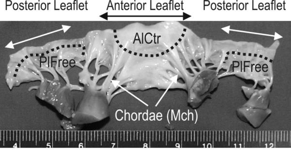
Mitral valve cut open to show ventricular aspect. Dotted lines and arrows denote the regions from which the three cell types were derived. AlCtr = anterior leaflet center, MCh = mitral chordae, PlFree = posterior leaflet free edge.
Official websites use .gov
A
.gov website belongs to an official
government organization in the United States.
Secure .gov websites use HTTPS
A lock (
) or https:// means you've safely
connected to the .gov website. Share sensitive
information only on official, secure websites.
Mitral valve cut open to show ventricular aspect. Dotted lines and arrows denote the regions from which the three cell types were derived. AlCtr = anterior leaflet center, MCh = mitral chordae, PlFree = posterior leaflet free edge.