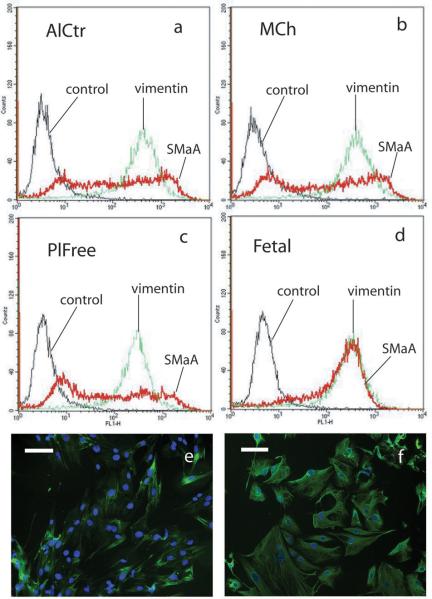
Figure 5.
(a-d) Representative histograms from fluorescence activated flow cytometry showing expression of SMαA and vimentin by AlCtr cells (a), MCh cells (b) and PlFree cells (c) as well as fetal porcine VICs (d). The fetal VICs were used in the flow cytometry as a positive control for SMαA. Negative controls were not incubated with the primary antibody. (e-f) Representative immunocytochemical stains showing SMαA in PlFree cells (e) and vimentin in AlCtr cells (f). Original magnification 10X.

