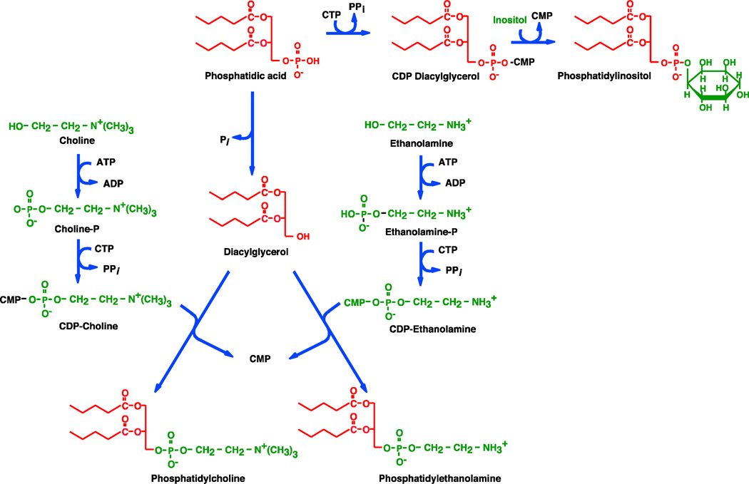
Figure 2.
Pathways for phospholipid biosynthesis in mammalian cells. The zig-zig lines in each red structure represent the long chain fatty acids on the phosphatidyl moiety. These are mostly saturated at the sn-1 position of unsaturated at the sn-2 position of the glycerol backbone. The components of each fatty acid are color coded as to the building blocks from which they are derived. The enzymes are located in the cytoplasm and the endoplasmic reticulum. The “Kennedy Pathway” generally refers to the pathways beginning with ethanolamine and choline. Not shown are the formation of PS from PE or PC by head group exchange with serine, the decarboxylation of PS to form PE, the methylation of PE by S-adenosyl methionine to form PC, and the formation of PG and CL in the mitochondria.

