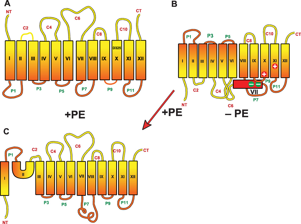
Figure 6.
Topological organization of LacY as a function of the presence or absence of PE. Cytoplasm is at the top of each figure, TMs are noted by rectangles, NT and CT refer to the N and C terminus respectively, and extramembrane domains oriented to the cytoplasm (C) or periplasm (P) in PE-containing cells are indicated. (A) LacY topology as determined in PE-containing (+PE) cells is depicted. The approximate position of D325 in TM X is indicated. (B) LacY topology after assembly in PE-lacking cells (−PE) cells. The exposure of TM VII (red) to the periplasm results in the loss of salt bridges of TM VII with TM X and TM XI as noted by the charges. (C) LacY topology determined after induction of PE synthesis in cells where assembly of LacY initially occurred in the absence of PE.

