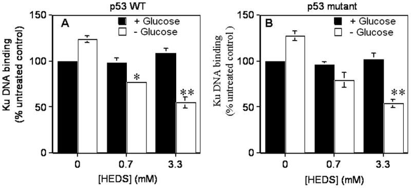
Figure 4. HEDS inhibits the function of redox-dependent DNA binding by Ku protein in glucose deprived cells.

Ku binding activity was compared after HEDS treatment of p53 wild type HCT116 (A) and p53 mutant HT29 (B) cells cultured in normal or glucose-deprived media. Mean for three to five independent experiments are shown with SD. A statistically significant reduction in Ku function in cells cultured in glucose-deprived media occurred after HEDS treatment (0.7 mM, *P<0.01; 3.3 mM, **P<0.001).
