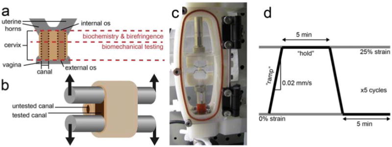
Figure 1.

Illustration of the rat cervix (a). The cervix was removed and cut in half, perpendicular to the canals. The section near the internal os was used for biochemical composition and collagen organization using birefringence measurements, while the section near the external os was used for biomechanical testing. The rat cervix has two canals, indicated by the vertical dotted lines, one leading to each uterine horn. For biomechanical testing, stainless steel rods were threaded through a single canal (b). A photograph of the cervix positioned in the custom-designed fixture attached to the Bose Biodynamic test system, submerged in phosphate buffered saline (c). The displacement waveform applied to each specimen, where 25% circumferential strain was calculated from the distance between the rods (d). Samples were stretched at 0.02 mm/s in the “ramp” section, then kept at 25% strain for 5 min during the “hold.”
