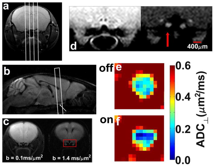
Figure 2.
Typical scout planning images (a,b), final targeted images for diffusion measurements (c,d), and ADC⊥ maps of an optic nerve without (e) and with visual stimulation (f). (a) A multi-slice set of axial images of the mouse brain is acquired with a non-diffusion-weighted multi-echo spin-echo image (only a single slice is shown here) are used to plan sagittal slices, indicated by the white lines. (b) The center sagittal slice, acquired with diffusion-weighting gradients applied in the slice-select direction, captures both optic nerves (arrow) and is used to target the slice for measurements of ADC⊥ and ADC||. (c) A pair of diffusion-weighted images with diffusion-sensitizing gradients applied in the phase-encode direction (left-to-right in the images). The optic nerve fibers are perpendicular to the slice plane in this final targeted slice. (d) Magnified view of the area within the red box of panel c. The left-hand image was acquired with b = 0.1 ms/μm2, and the right-hand image with b = 1.4 ms/μm2. Both optic nerves are clearly visible in the high-b image and the stimulated optic nerve indicated by the red arrow. Color maps of ADC⊥ in an experimental optic nerve without (e) or with (f) visual stimulation.

