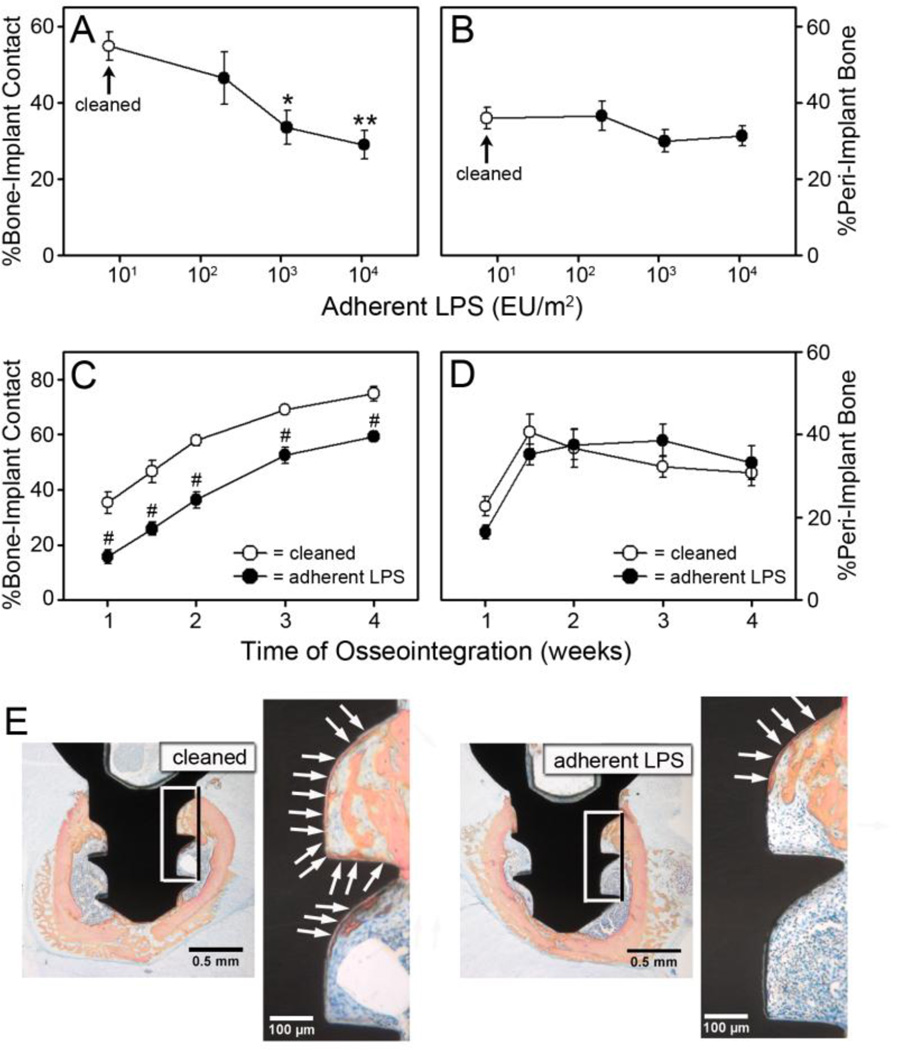
Figure 2. Adherent LPS inhibits bone-to-implant contact.
The percentage of bone-to-implant contact (A) and the percentage of peri-implant bone (B) at one week after implantation in wild-type C57Bl/6J mice with rigorously cleaned implants (open circles) or implants with adherent LPS (closed circles). The percentage of bone-to-implant contact (C) and the percentage of peri-implant bone (D) over a four week time course in wild-type C57Bl/6J mice with rigorously cleaned implants (open circles) or implants with 1 × 10⁴ EU/m2 of adherent LPS (closed circles). Representative histological cross-sections, which are closest to the mean in C, at one week after implantation in mice with rigorously cleaned implants or implants with 1 × 10⁴ EU/m2 of adherent LPS (E). Boxes indicated area magnified in next panel. Arrows denote bone-to-implant contact. Statistical analysis was by One-Way ANOVA (A and B) or by Two-Way ANOVA (C and D). * denotes p=0.017, ** denotes p=0.003, and # denotes p<0.001 compared to rigorously cleaned. n=8–11.

