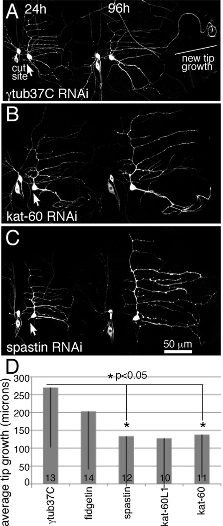
Figure 1. Regeneration of an axon from a dendrite is sensitive to levels of microtubule severing proteins.
EB1-GFP, dicer2 and hairpin RNAs were expressed in class I sensory neurons with 221-Gal4. The axon of the ddaE neuron was severed close to the cell body with a pulsed UV laser at 0h. Animals were remounted for imaging at 24h intervals. The 24h and 96h timepoints are shown. At 24h the axon is completely gone. A. At 96h in control neurons expressing a hairpin targeting γtub37C, a neurite can be seen to extend from beyond the normal territory covered by the dendrite. Comparison of the 24h and 96h images makes it clear that one of the dendrite tips has grown between the two time points. B and C. When either kat-60 or spastin was targeted by RNAi many cells did not extend their dendrites as in these examples. D. The length of outgrowth from dendrite tips was measured in neurons expressing different hairpin RNAs. Numbers in the columns indicate the number of animals of each genotype tested. Error bars show standard deviation; as these were fairly large the bar is shown in only one direction. Only part of the error bar is shown for kat-60L1; the standard deviation was 181 microns. Statistical significance was calculated with a student’s t-test.

