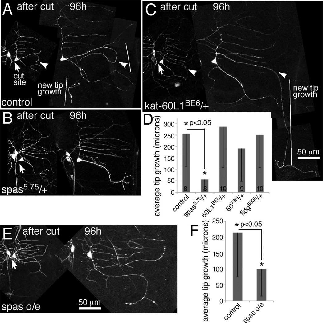
Figure 2. Too little or too much spastin reduces regeneration of an axon from a dendrite.
Proximal axotomy was performed as in Figure 1. A–C. Images from animals immediately after ddaE axon severing are shown together with the same cell 96 hours later. Arrows indicate the site of severing, and arrowheads in A and C point to the dendrite that initiates tip growth. D. The length of new growth from a dendrite tip was quantitated. Numbers in the columns indicate the number of animals of each genotype tested. Error bars show the standard deviation, and this is shown in one direction only to keep the graph compact. Statistical significance was calculated with a t-test. E and F. Neurons expressing EB1-GFP and either mCD8-RFP (control) or spastin-CFP (spas o/e) were subjected to proximal axotomy. Tip growth at 96h after injury was quantitated as in D, error bars and significance are also the same as in D. An example of a spastin overexpressing ddaE cell is shown in E.

