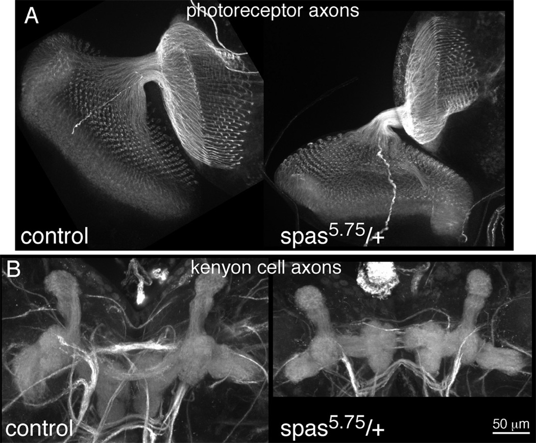
Figure 7. Axon outgrowth is normal in spastin heterozygotes.
Axons of photoreceptor neurons and kenyon cells were stained in dissected larval brains. A. Axon projections from the developing eye (left/ bottom) to the brain (top/ right) are shown. B. Axon projections of kenyon cells in the central brain are shown. Dorsal axon projections point to the top of the image, and medial projections are the tri-lobed structures pointing towards the middle of the image. Axons were stained with a cocktail of monoclonal antibodies recognizing Dac, FasII and Trio.

