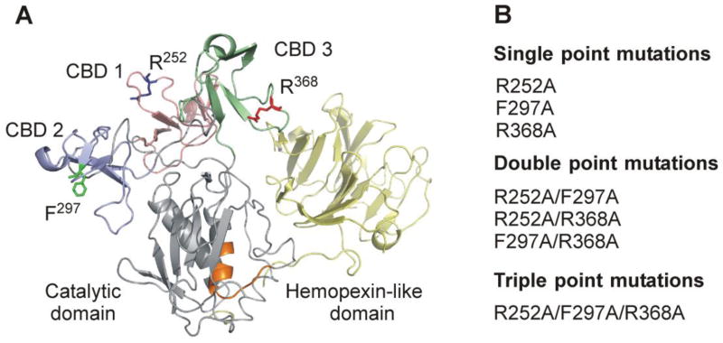
Fig. 1.
Structural overview of MMP-2 highlighting the locations of modified collagen binding site residues. (A) Crystal structure of MMP-2 (PDB 1CK7) (Morgunova et al., 1999) with modified collagen binding site residues (R252, F297, R368) presented in stick form (PyMOL). The three fibronectin type II modules of the collagen binding domain are shown in pink, blue, and green, respectively, and the enzyme active site is depicted in orange. (B) Alanine substitutions were introduced into one or concurrently into two or three of the CBD modules in full-length MMP-2 by a PCR-based site-directed mutagenesis strategy.

