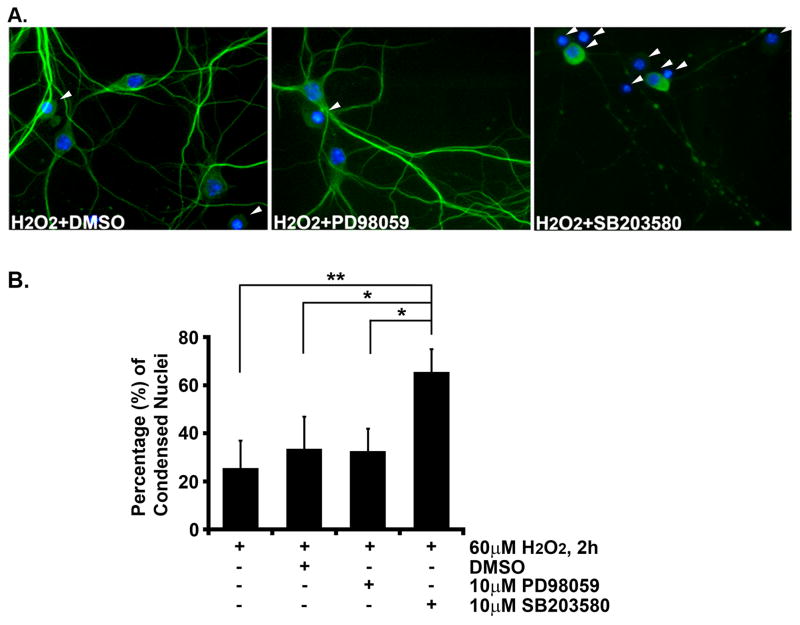
Fig. 3. Inhibition of p38 MAPK but not ERK blocks RitQ79L-mediated protection from reactive oxygen species-induced death.
(A) Hippocampal neurons (DIV8) were pre-treated for 30 min with DMSO, PD98059, or SB203580, and subjected to H2O2 exposure (60 μM) for 2 h. Cells were fixed, stained (neurons, MAP2, green; nuclei, Hoechst, blue) and scored for condensed nuclei (arrowhead). Representative images are shown. (B) The percentage of MAP2+ cells with condensed nuclei are presented as mean ± SEM. Experiments were performed in triplicate. * p<0.02, ** p<0.006 using Fisher’s PLSD post-hoc test.

