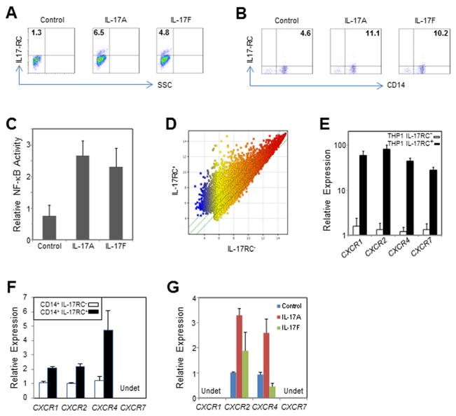
Figure 3. Induction of IL-17RC by IL-17A and IL-17F in monocytes and characteristics of IL-17RC+ monocytes. (Data are expressed as the means ± S.E.).
(A) FACS staining of IL-17RC induced by IL-17A and IL-17F in THP1 cells.
(B) FACS staining of IL-17RC induced by IL-17A and IL-17F in primary CD14+ monocytes. See also Figure S3.
(C) Relative activity of NF-κB measured by TransAM NF-κB p65 Transcription Factor ELISA Kit.
(D) Scatter plot showing the genome-wide expression difference between sorted IL-17RC+ and IL-17RC− THP1 cells.
(E) Relative expression of CXCR1, CXCR2, CXCR4, and CXCR7 mRNA in sorted IL17RC+ and IL17RC− THP1 cells.
(F) Relative expression of CXCR1, CXCR2, CXCR4, and CXCR7 mRNA in sorted IL17RC+CD14+ and IL17RC−CD14+ monocytes from two AMD patients. Undet, undetectable.
(G) Relative expression of CXCR1, CXCR2, CXCR4, and CXCR7 mRNA in primary CD14+ monocytes stimulated by IL-17A or IL-17F overnight. Undet, undetectable.

