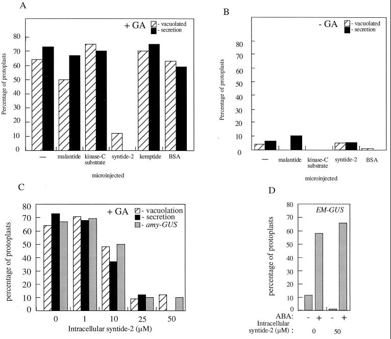
Figure 1.
The effect of microinjection of protein kinase peptide substrates on GA-induced amylase secretion, vacuolation, and amylase-GUS and EM-GUS expression. A and B, Freshly isolated protoplasts were microinjected with 50 μm of the indicated protein kinase substrate peptides or BSA and then treated for 24 h with (+GA) or without (−GA) 5 μm GA. C, Freshly isolated protoplasts were microinjected with a range of syntide-2 concentrations and then treated for 24 h with 5 μm GA. Secretion of amylase, development of GA-induced vacuolated morphology, and amylase-GUS expression were then assessed on a single-cell basis. D, Freshly isolated protoplasts were co-microinjected with 50 μm syntide-2 or BSA and the EM-GUS construct and treated with or without 10 μm ABA. Induction of EM-GUS expression was then assessed on a single-cell basis. Protoplasts were scored as showing induction of amylase-GUS or EM-GUS expression if they exhibited a GUS:LUC of more than 4000 (Gilroy, 1996). Protoplasts were scored as secreting amylase if they showed a cleared “halo” of >25 μm in the starch thin-film assay (Hillmer et al., 1992). Vacuolated protoplasts were those showing development to stage 3 or 4 as defined by Bush et al. (1986). The responses of at least nine microinjected protoplasts per treatment are shown.

