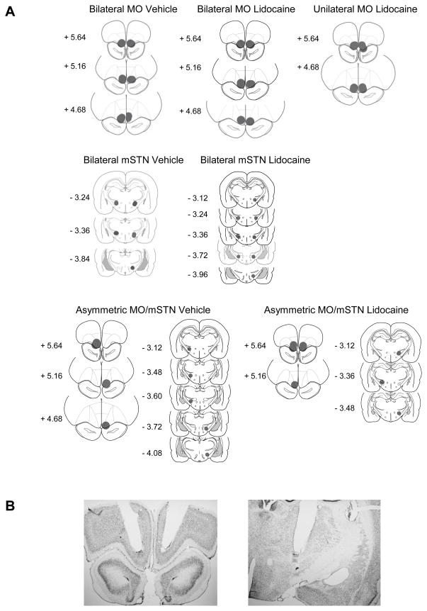
Figure 2.
(A) Schematic drawings representing coronal sections depicting placements in medial orbital cortex (MO) and medial tip of the subthalamic nucleus (mSTN). Circles (0.5 mm radius, drawn to scale) indicate the location and theoretical spread of infused drug, based on the spherical volume equation for lidocaine [31]. In all drawings, the anterior-posterior (AP) reference points are measured in mm from bregma, and each placement is shown at the midpoint of its AP extent. (B) Representative low-magnification (2X) photographs of guide cannulae placements terminating 1 mm above the MO (left) and mSTN (right).

