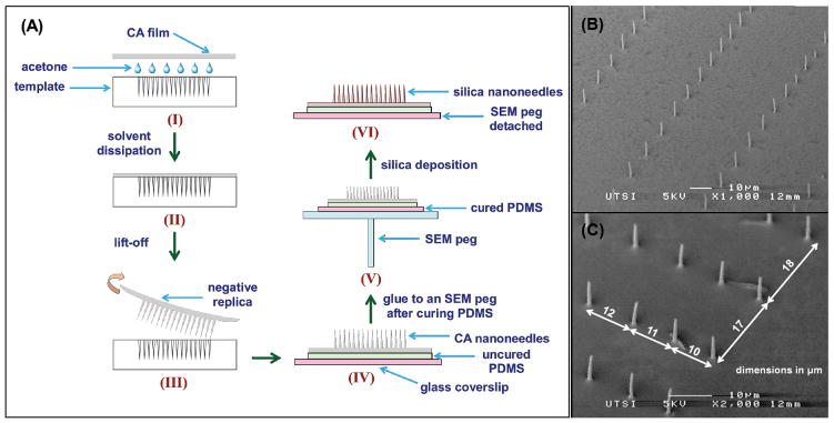
Figure 1. Pattern prepared on fused silica substrate by single-pulse femtosecond laser machining.
(A) A schematic of the pattern displaying the 2D gradient in nanohole spacing (nanohole density reduced for visual clarity). The pattern is a 2×2 matrix of four quadrants, each formed by increasing the spacing between successive nanoholes by 1 μm starting from 10 μm in the densest location (edge) to 50 μm in the sparsest location (middle of the quadrant) and then decreasing from 50 μm to 10 μm at 1 μm decrements in both x and y directions. A quadrant is a matrix of 84×84 nanoholes, and a 2×2 matrix of these quadrants forms the pattern with 168×168 nanoholes. Each black dot in the schematic represents a nanohole made by focusing a single laser pulse. (B) An SEM image of the nanoholes from one edge of the pattern.

