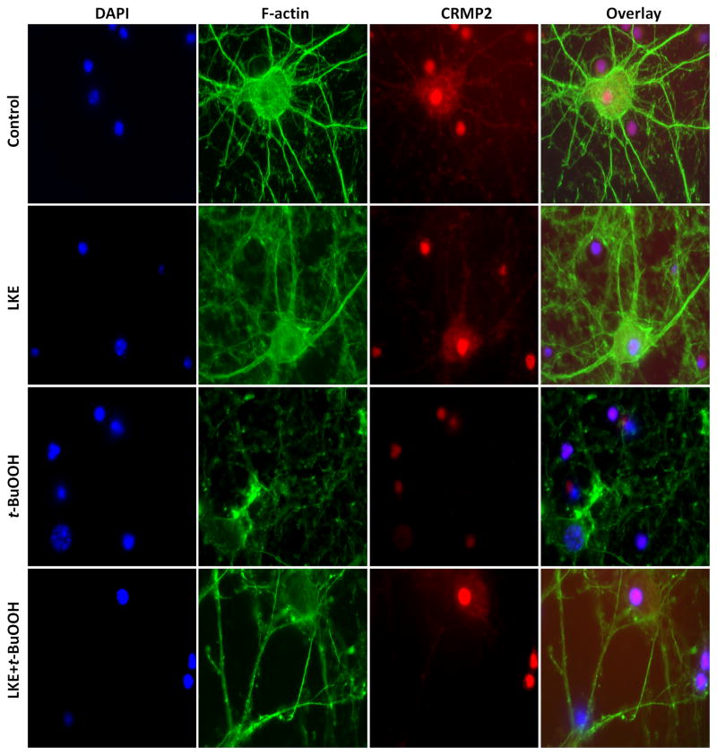
Figure 4.
LKE elicits neuritogenesis in mice cortical neurons. Top panel: control neurons without any treatment. The nucleus is stained with DAPI (blue stain), F-actin (phalloidin green stain), and CRMP2 (red stain). The far right image shows the merged overlay of all the pictures. t-BuOOH treatment induced CRMP2 localization into the nucleus and collapse of F-actin filaments. Pre-treatment with LKE (200 μmol/L) induced the expression of CRMP2 and F-actin in axons and dendrites. Each experiment was conducted in triplicate and repeated three times with different primary culture batches.

