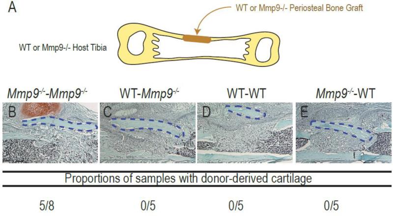
Fig. 3. Analysis of cartilage formation during healing of Mmp9–/– and wild type periosteal bone grafts.
(A) Experimental design. (B-E) Longitudinal sections through the tibia and histological evaluation using Safranin-O staining at day 10 post-surgery of Mmp9–/– periosteal bone grafts transplanted in Mmp9–/– (B, Mmp9–/– - Mmp9–/–) or wild type hosts (C, Mmp9–/–-WT) or wild type periosteal bone grafts transplanted in wild type (D, WT-WT) or Mmp9–/– hosts (D, WT-Mmp9–/–). Cartilage formation is only observed in Mmp9–/– mice transplanted with Mmp9–/– bone grafts. Student's t-test: * P<0.05 (Mmp9–/–-Mmp9–/–compared to WT-Mmp9–/–, WT-WT and Mmp9–/–-WT; n=5 or 6 per group).

