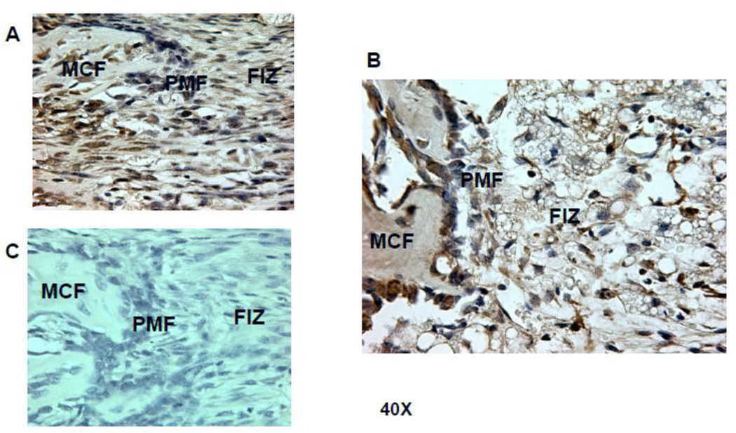
Figure 8.
Osteocalcin stain of PMF region (brown) highlights osteoblasts, osteocytes with matrix and bone matrix at 40X magnification. A. The PMF in an untreated Avy/a mouse demonstrates obvious osteoblasts densely lining and within the new trabeculae but not extending into the FIZ. B. The PMF in a Rosi treated Avy/a mouse showing a smaller blunted new bone trabeculum and isolated osteocalcin positive cells within a disorganized FIZ infiltrated with fat cells as identified by aP2 stain in figure 6. C. The PMF in an untreated Avy/a mouse stained with normal rabbit IgG instead of the primary antibody as a control. D. Quantification of OC-positive cells by cell counting within the MCF region of interest, comparing the four groups(a/a, Avy/a, Untreated “C”, Rosi treated “R”). p > 0.05.

