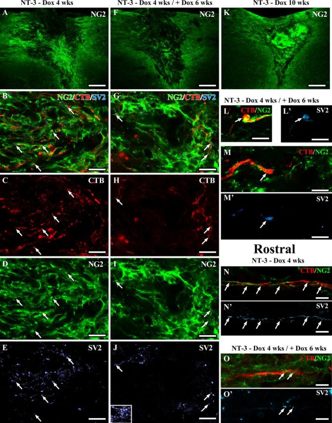
Figure 13.

Regenerated sensory axons associate with NG2+ glia and form synapse-like contacts. Confocal images of sections triple immunolabeled for CTB-labeled sensory axons (red), NG2+ cells (green), and the presynaptic marker SV2 (light blue). Low-magnification overview of NG2 labeling in and around the lesion site shows higher NG2 labeling in the graft of animals with (A) 4 weeks or (K) 10 weeks of NT-3 expression compared with animals with (F) transient NT-3 expression (six weeks after reduction of NT-3 levels). Numerous regenerated axons are closely associated with NG2+ cells in (B–M) the graft and in (N–O) the rostral white matter. NG2 and CTB- and SV2-labeling density is higher in (A–E) grafts of animals with continuous NT-3 expression for 4 weeks compared with(F–J) animals with transient NT-3 expression. Inset in J shows SV2 labeling in gray matter as positive control. Arrows indicate SV2 labeling colocalizing with CTB-labeled axons adjacent to NG2+ glia. L, M, Six weeks after reduction of NT-3 expression, SV2-labeling is particularly strong in CTB-labeled axons with an end bulb-like appearance surrounded by NG2+ glia. SV2 labeling is also present in (N, O) regenerated CTB-labeled sensory axons associated with NG2+ glia rostral to the lesion site. Scale bars: A, F, K, 226 μm; B–E, G–M, 10 μm; N–O, 5 μm.
