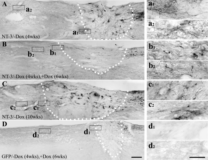
Figure 8.
Immunolabeling for regenerated CTB-labeled sensory axons after transient NT-3 expression. Many regenerating axons are found beyond the lesion site when NT-3 gene expression is turned on (−Dox) for (A) 4 weeks and (C) 10 weeks. B, Fewer CTB-labeled regenerated axons are observed in the graft and in the rostral spinal cord when NT-3 expression is turned on (−Dox) for 4 weeks and then turned off (+Dox) for 6 weeks. D, As expected, very few CTB-labeled axons extend into the graft and beyond the graft in animals received transient GFP gene delivery. Dotted lines indicate the host/graft interface. Higher magnification of boxed areas in A–D are shown in a1–d1 (rostral host/graft interface) and a-2–d2 (rostral to the lesion site). Scale bars: (in D) A–D, 200 μm; (in d2) a1--d2, 50 μm.

