Fig. 1.
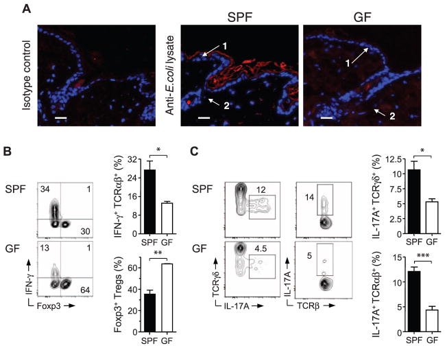
Commensal microbiota control the balance of effector and regulatory T cells in the skin tissue. (A) Immunofluorescence labeling of bacterial products in interfollicular keratinocytes (1) and hair follicles (2) from skin tissue of SPF and GF mice. Representative images show naïve skin stained with anti–E. coli lysate antibody (red) or isotype control and Hoechst (blue); scale bars, 25 μm. (B) Representative flow cytometric plots and summarized bar graphs of IFN-γ and Foxp3 expression by live CD45+ TCRβ+ cells extracted from skin tissue of SPF and GF mice after stimulation with phorbol myristate acetate (PMA) and ionomycin. Graphs show means ± SEM of three or four mice (*P < 0.05, **P < 0.005). Results are representative of three experiments. (C) Representative flow cytometric plots and summarized bar graphs of IL-17A expression in live CD45+ TCRγδ+ or CD45+ TCRαβ+ cells from skin tissue of SPF and GF mice after stimulation with PMA and ionomycin. Graphs show means ± SEM of three or four mice (*P < 0.05, ***P < 0.0005). Results are representative of three experiments.

