Fig. 3.
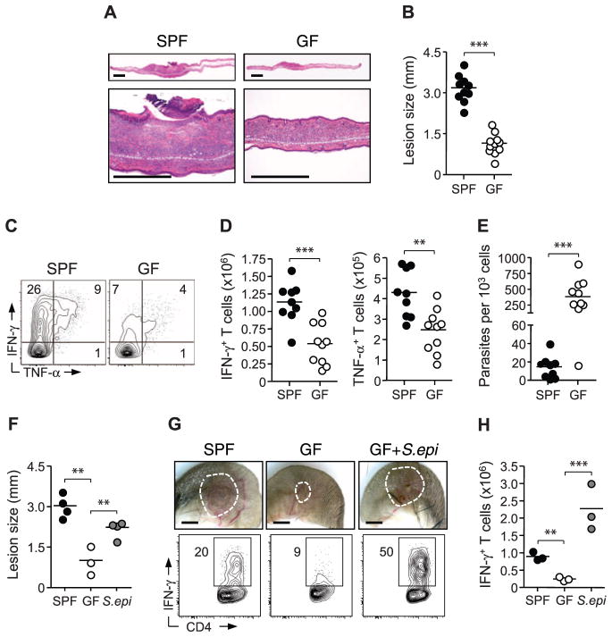
Cutaneous commensals drive immunity and promote pathology in L. major infection. (A) Histopathological comparison of ear pinnae skin lesions from L. major–infected SPF and GF mice. Scale bars, 500 μm. (B) Assessment of lesion size in SPF and GF mice. Each data point represents an individual mouse (***P < 0.0005). (C and D) Flow cytometric analysis of Leishmania antigen-specific IFN-γ and TNF-α production by TCRβ+ CD4+ dermal cells from L. major–infected SPF and GF mice. Each data point represents an individual mouse (**P < 0.005, ***P < 0.0005). Results are representative of three experiments. (E) Number of L. major parasites per 1000 nucleated cells from dermal lesions of infected SPF and GF mice. Each data point represents an individual mouse (***P < 0.0005). (F) Assessment of lesion size in SPF mice, GF mice, and GF mice monoassociated with S. epidermidis (S.epi). Each data point represents an individual mouse (**P < 0.005). Results are representative of two experiments. (G and H) Representative images of L. major skin lesions and analysis of IFN-γ production by live TCRβ+ CD4+ cells from SPF mice, GF mice, and GF mice mono-associated with S. epidermidis. Each data point represents an individual mouse (***P < 0.0005). Results are representative of two experiments.

