Figure 2.

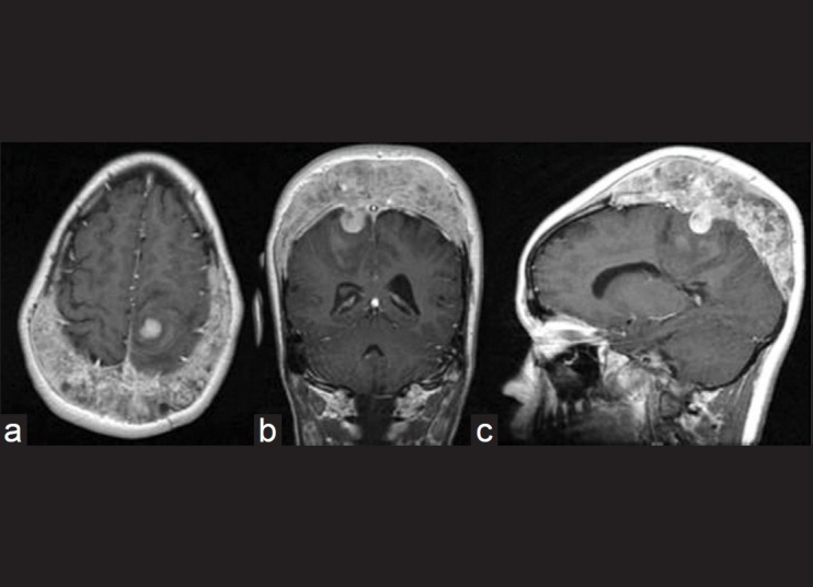
Posthemorrhage (2010) axial (a), coronal (b), and sagittal (c) T1-weighted postcontrast magnetic resonance imaging showing a posterior frontoparietal parenchymal hematoma, along with progression in tumor size

Posthemorrhage (2010) axial (a), coronal (b), and sagittal (c) T1-weighted postcontrast magnetic resonance imaging showing a posterior frontoparietal parenchymal hematoma, along with progression in tumor size