
Figure 1.
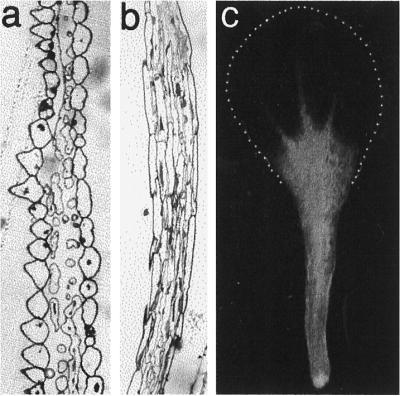
Structure of mature Arabidopsis petals. a, Transverse section through the upper white petal lamina. b, Transverse section through the stalk at the basal part of the petal. c, Entire intact petal viewed by conventional fluorescence microscopy showing chlorophyll autofluorescence in the petal stalk and the lack of fluorescence in the upper white petal lamina. The extent of the white petal lamina is shown by the dotted line.