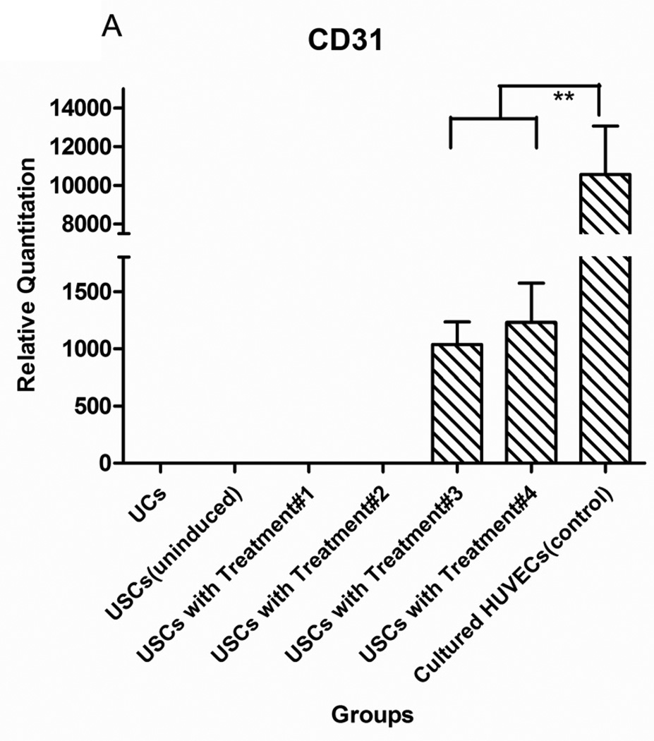
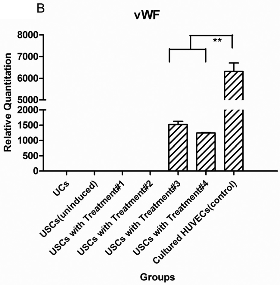
Fig. 2. Endothelial gene expression of USCs in vitro.
USC (P3) were seeded at 1,000 cells/cm2 and induced by endothelial differentiation media as follows: Treatment#1= DMEM (10%FBS) with VEGF alginate beads located in transwell; Treatment#2= DMEM (10%FBS+1%P/S); Treatment#3=EC induced medium (EGM-2) plus alginate microbeads loaded with VEGF; Treatment#4= Endothelial cell-induced medium including VEGF solution (10 ng/ml). Significant increase of endothelial cell-specific gene expression CD31 (A) and vWF (B) could be detected in both Treatments #3 and 4, regardless of whether VEGF was added directly to the medium or delivered by the alginate beads. Urothelial cells (UC) were the negative control and human umbilical vein endothelial cells (HUVEC) were the positive control. **p<0.01.


