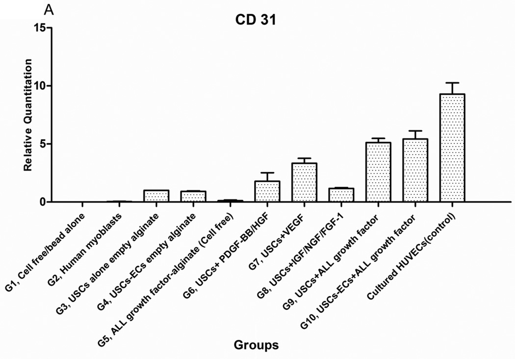
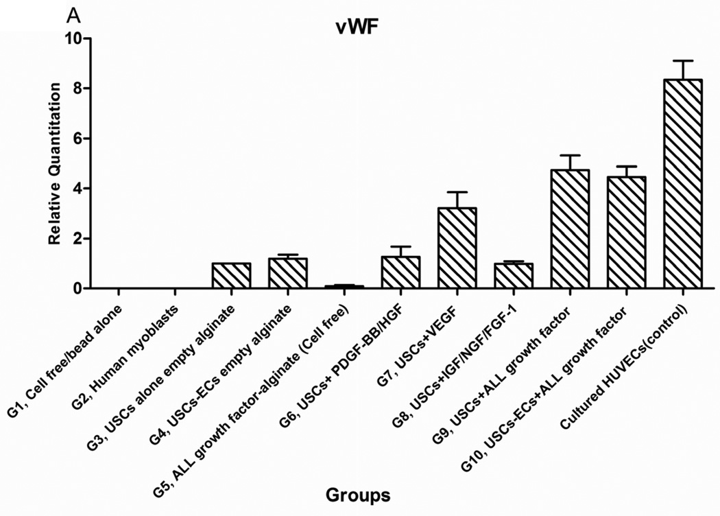
Fig. 5. Endothelial differentiation of USCs and angiogenesis of implanted grafts 28 days after implantation in vivo.
A) Quantitative real-time PCR was performed on total RNA from all groups using endothelial-specific primers (CD31, vWF). B) Groups 1, 3, 4, 5, 7, and 9 were subjected to immunofluorescent staining using the epithelial markers CD31 and von Willebrand factor (vWF) with Human nuclei specific marker. Specific staining (shown by arrows) appears green with nuclear staining in red (human nuclei) and blue (DAPI) indicating these were implanted cells that successfully differentiated to endothelial cells in vivo. Scale bar = 50 µm.


