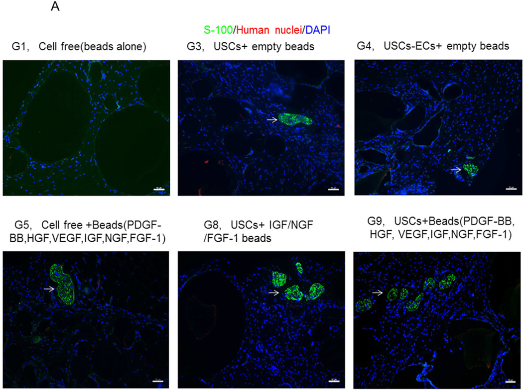
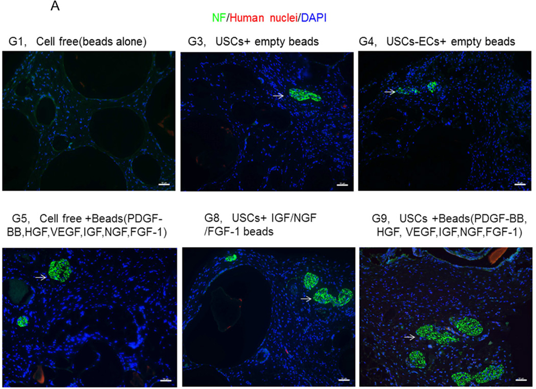
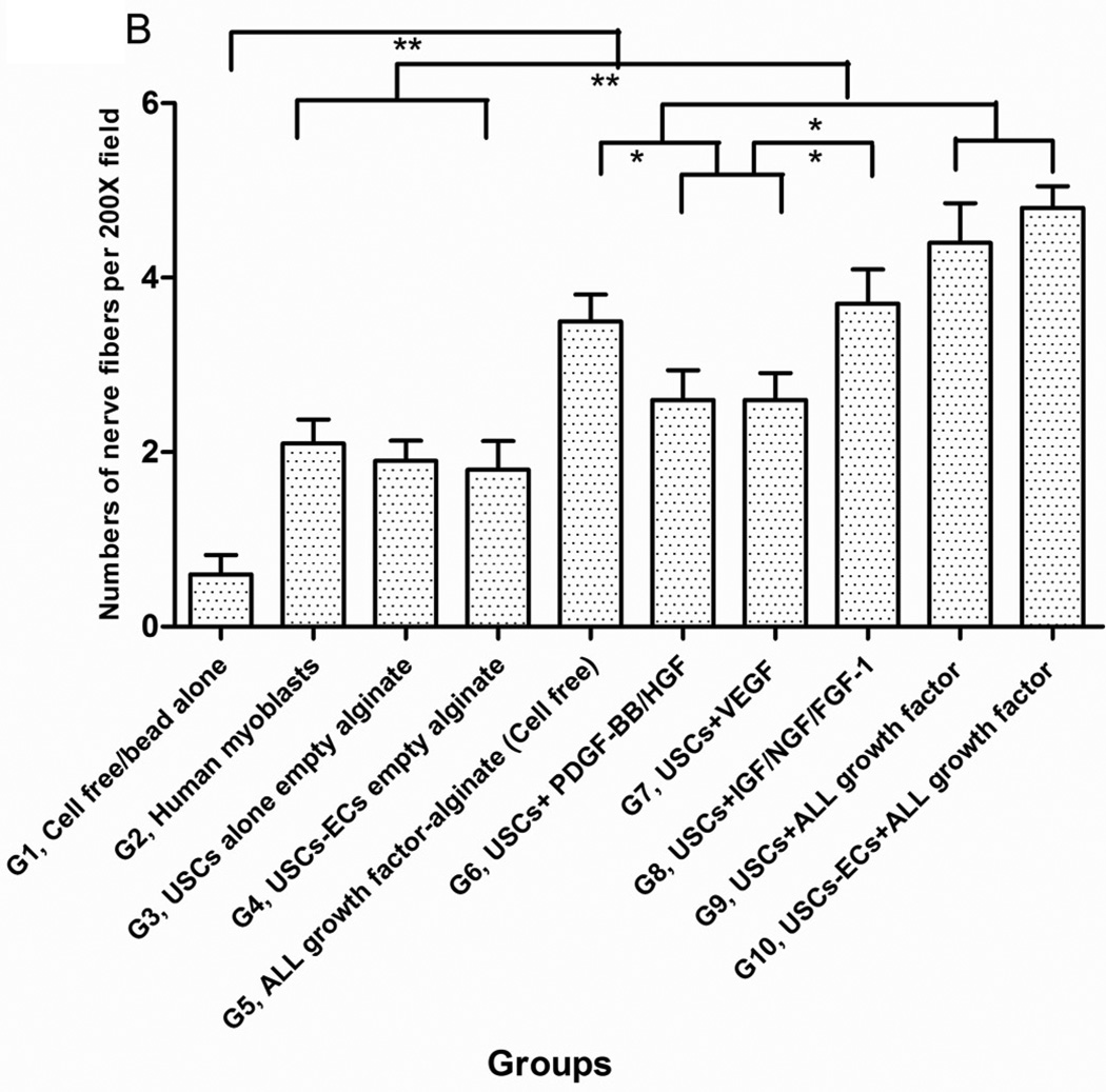
Fig. 6. Innervation of implanted grafts 28 days after implantation.
A) Cross-sections of samples from Groups 1, 3, 4, 5, 8, and 9 were subjected to immunofluorescent staining using DAPI (blue), human nuclear (red) and nerve cell antibodies (green)-S-100 and Neurofilament (NF). Scale bar= 50 µm. B) Semiquantitative analyses of nerve fibers in the implanted grafts. *p<0.05, **p<0.01.


