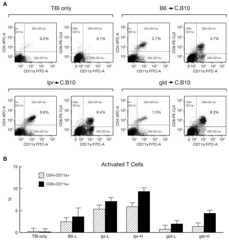
FIGURE 5.
Expansion and activation of T cells. Residual BM cells from mice discussed in Figure 4 were stained with an antibody mixture containing CD4-APC, CD8-PeCY5 and CD11a-FITC. The majority of CD4+ and CD8+ T cells were positively stained for the activation marker CD11a in mice that received LN cell infusions (A). CD4+ and CD8+ T cell proportions were significantly higher in mice that received lpr LN cells (B).

