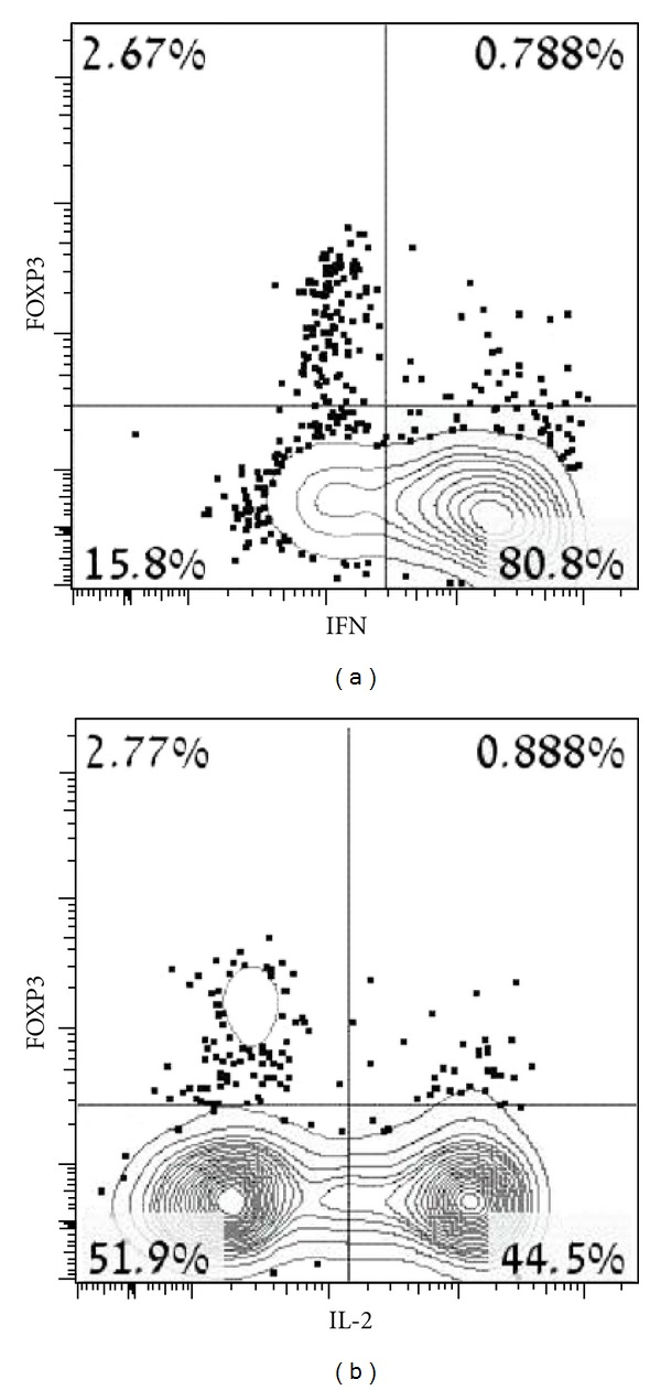
Figure 5.

IFNγ and IL-2 cytokines secretion following T-cell stimulation. PBMCs obtained from patient 2 were stimulated with PMA and ionomycin, than stained with CD4, FOXP3, and IFNγ or IL-2 for the identification of functional Tregs. Detection was performed using FACS analyses. Quadrants were set up based on staining with isotype control. Boxed numbers indicate the percentage of cells within the CD4+ population that secrete IFNγ or IL-2.
