Fig. 5.
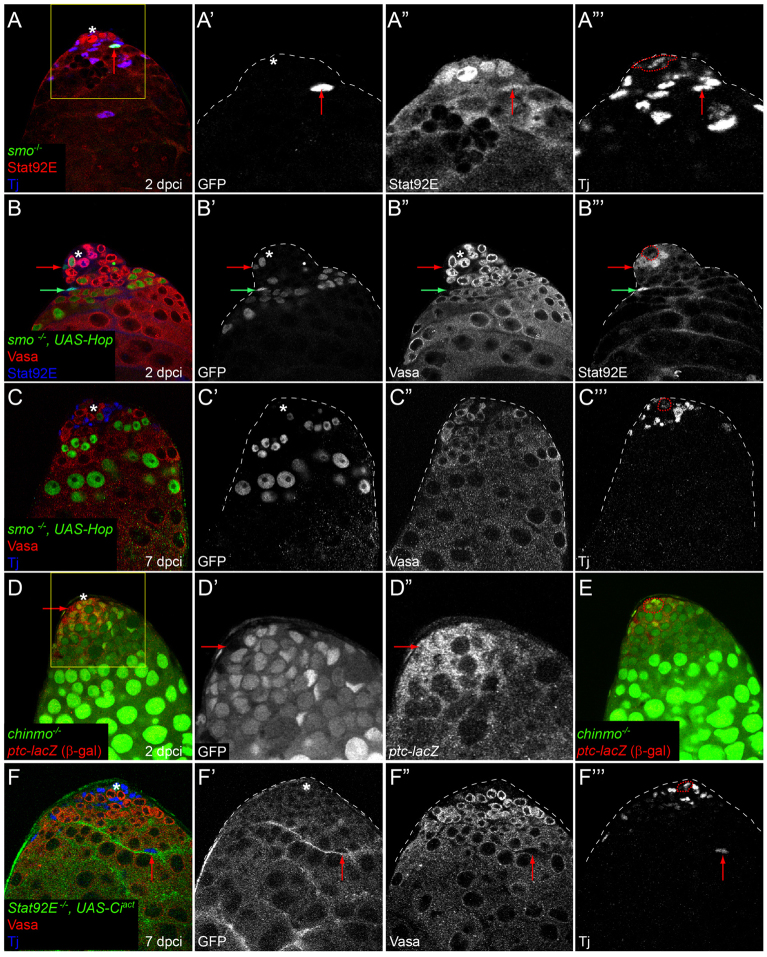
Relationship between Hh and JAK/STAT signaling in CySC self-renewal. (A-A″′) No change in Stat92E expression is detected in positively labeled smo mutant cells (A,A″′; arrow indicates smo mutant clone) at 2 dpci. Boxed area in A is enlarged in A′-A″′. GFP is green (single channel in A′), Stat92E is red (single channel in A″) and Tj is blue (single channel in A″′). (B-B″′) Stat92E is stabilized in smo MARCM clones that express Hop. Red arrow indicates a smo mutant CySC and the green arrow indicates a differentiating cyst cell. GFP is green (single channel in B′), Vasa is red (single channel in B″) and Stat92E is blue (single channel in B″′). (C-C″′) Activation of Stat92E by overexpression of Hop cannot rescue loss of smo MARCM mutants from the stem cell niche at 7 dpci, despite the stabilization of Stat92E protein in these clones (see B,B″′). GFP is green (single channel in C′), Vasa is red (single channel in C″) and Tj is blue (single channel in C″′). (D-D″) ptc-lacZ (red; single channel in D″) expression is unaltered in negatively marked chinmo mutants (arrow points to a ptc-lacZ-expressing mutant CySC in D-D″). Boxed area in D is enlarged in single channels in D′,D″. chinmo clones lack GFP (single channel in D′). (E) Adjacent optical z-section to D, showing where the hub (red dotted outline) is located. (F-F″′) Ciact expression cannot rescue Stat92E MARCM mutant CySCs at 7 dpci, but differentiated cyst cells can be recovered (red arrow indicates marked cyst cell; clones were labeled with membrane-targeted GFP). See also Table 1. The position of the hub is indicated with an asterisk or outlined in the Tj single channel.

