Fig. 3.
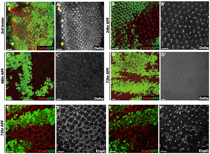
Delta-independent activation of Notch signaling in Abl clones. (A-D′) Delta expression in third instar (A), 24 hours APF (B), 48 hours APF (C) and 72 hours APF (D) eye disks. All pictures are confocal maximal projections. The MF is marked by yellow arrows in A. Delta protein is normally expressed in Abl clones, with its level decreasing gradually during development. At 72 hours APF, when Notch is highly expressed in Abl clones, no ectopic Delta protein is detected. (E,E′) Notch signaling is elevated as assessed by the increase in E(Spl)+ cells (red) in Abl clones. Maximal projection. (F,F′) A basal section of E showing the extra E(spl)+ cells reside at the basal plane. Scale bars: 10 μm.

