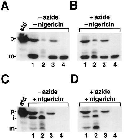
Figure 4.
A, Reactions incubated in the absence of both inhibitors; B, reactions incubated with azide alone; C, reactions incubated with nigericin alone; and D, reactions incubated with both inhibitors. Lanes 1, Intact chloroplasts; lanes 2, stromal fractions; lanes 3, washed thylakoids; and lanes 4, protease-treated thylakoids. A 4× import reaction was initiated by the addition of 80 μg of chlorophyll and a 30-min import reaction was conducted in the light. At this time, one aliquot was removed and the sample was centrifuged through silicon oil into PCA; these intact chloroplasts were run in lanes 1. The remaining chloroplasts were pelleted for 30 s in a microfuge, the supernatant was removed, and the chloroplasts were resuspended in 0.5 volume of envelope lysis buffer containing 4 μg of aprotinin, 4 μg of leupeptin, and 1 mm PMSF. To distinguish between mOE17 bound to the lumenal face of the membrane and proteins that were fully translocated, we protease treated one-half of the thylakoids with thermolysin (lanes 4), whereas the other half was mock-protease treated (lanes 3). The thylakoid fraction also contained fragmented envelope membranes; thus, some prOE17 is seen in the samples that are not protease treated (lanes 3). The standard (std) represents 20% of the precursor added to each reaction; the positions of the precursor (p), intermediate (i), and mature (m) proteins are noted.

