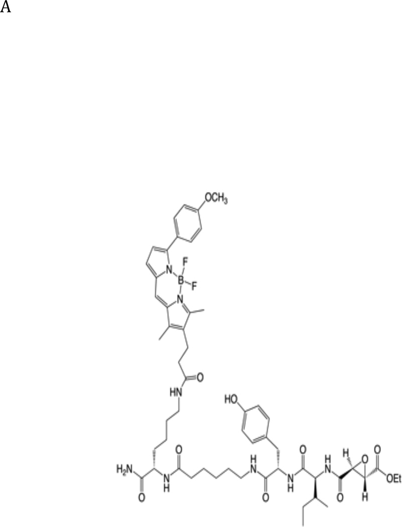
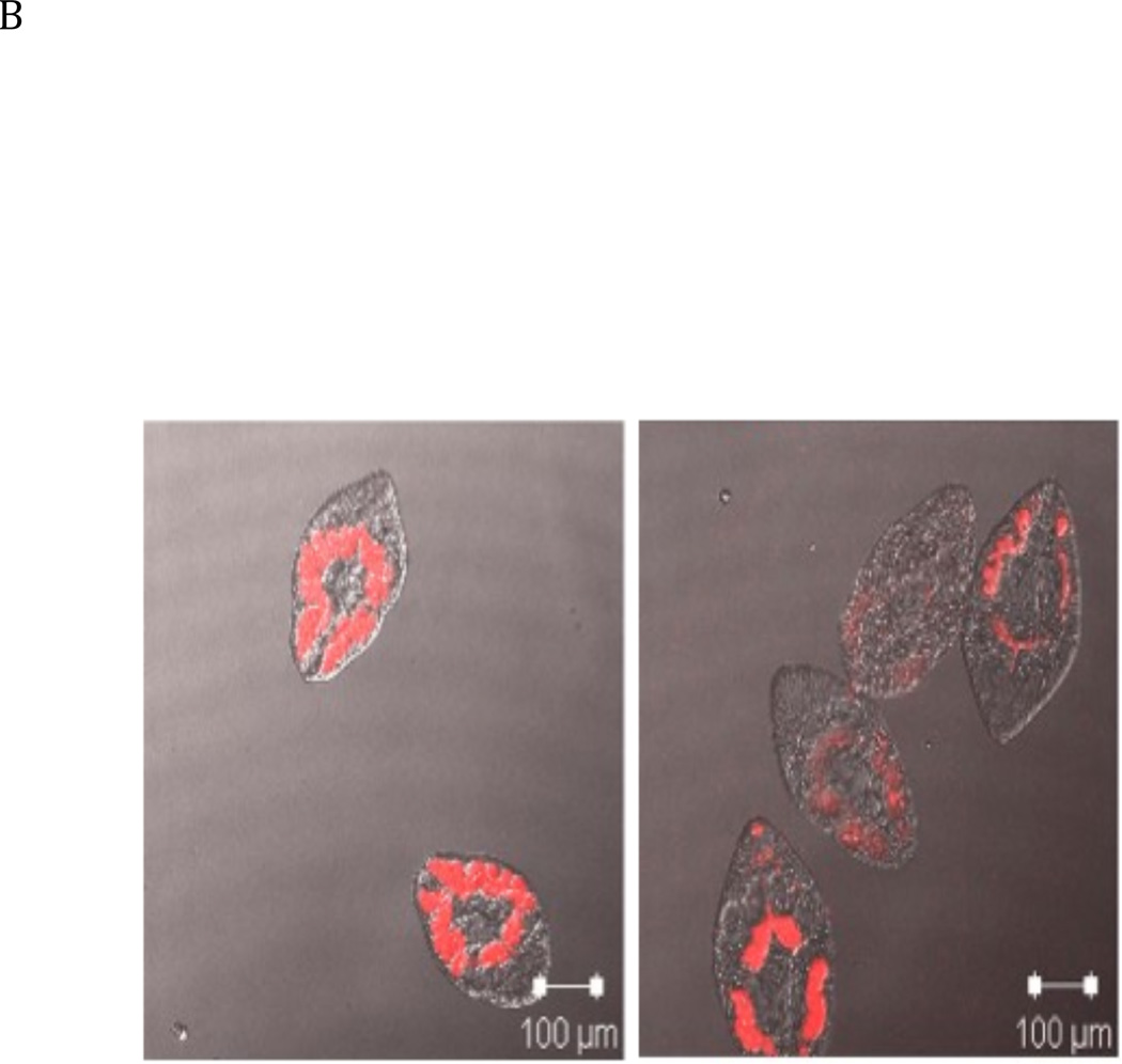
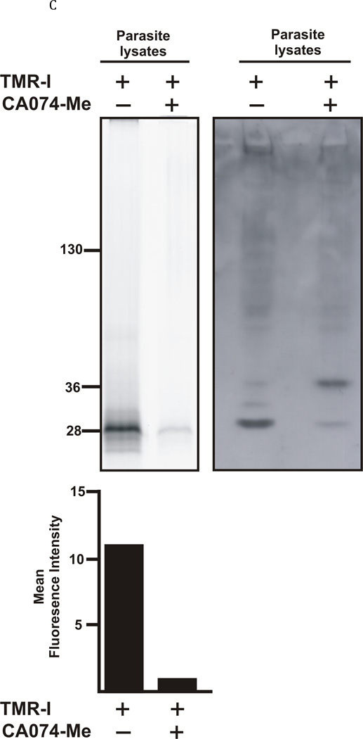
Fig. 5. Incubation of Fasciola hepatica juvenile flukes with a fluorescent active site probe reveals the localisation of active cathepsin B like enzymes.
A: Structure of the TMR-I probe used for visualization of the cathepsin B enzymes within the parasites. B: 40 newly excysted juvenile parasites per well were either exposed to the TMR-I (6.25 µM) for 24 hrs (left panel) or exposed to CA074Me (6.25 µM) for 7 days prior to exposure to the TMR-I (6.25 µM) for 24 hrs (right panel). C: Determination of the molecular target of TMR-I and CA074Me. The left hand panel shows a fluorogram of the binding of the TMR-I to proteins in a lysate from F. hepatica NEJ flukes. As indicated, proteins were incubated with the TMR-I in the presence and absence of CA074Me. The intensity of the bands obtained with and without CA074Me is shown below the fluorogram. The right hand panel shows an immunoblot with rabbit anti-FhcatB1 against parasite lysate proteins treated with TMR-I in the presence and absence of CA074Me.


