Figure 5.
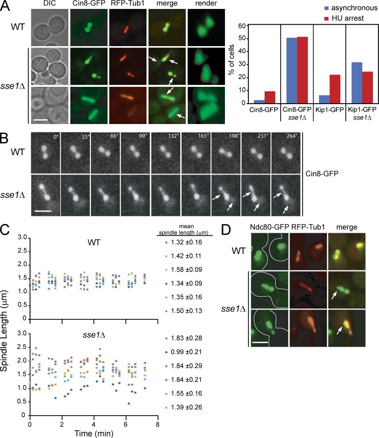
Localization of GFP-tagged kinesin-5 motors in WT and sse1Δ cells. (A) WT and sse1Δ cells expressing RFP-Tub1 and Cin8-GFP were synchronized in S phase with HU for 2.5 h at 26°C. Representative images (extended focus and 3D render) of Cin8-GFP (green) and RFP-Tub1 (red) obtained using fluorescence confocal microscopy are shown. Arrows point to the presence of Cin8 in the midzone and nucleoplasm. Bar, 5 µm. The bar graph shows the percentage of cells of the indicated genotype that have asymmetric distribution of Cin8 in either asynchronous or HU-arrested cultures. About 200 cells were observed. The data shown are from a single representative experiment out of three repeats. DIC, differential interference contrast. (B) WT and sse1Δ cells expressing Cin8-GFP were incubated with HU for 2.5 h, and time-lapse microscopy was performed. The images show that deletion of SSE1 results in redistribution of Cin8 localization and, hence, in varied spindle length. The arrows show the presence of Cin8 in the midzone and nucleoplasm. Bar, 5 µm. (C) Spindle length is plotted for six HU-arrested WT or sse1Δ cells as a function of time. The mean spindle length measured for each cell over the indicated time period is given on the right. (D) WT and sse1Δ cells expressing Ndc80-GFP (green) and RFP-Tub1 (red) were synchronized in S phase with HU for 2.5 h at 26°C. Images were obtained using fluorescence confocal microscopy. Arrows point to the tighter clustering of Ndc80-GFP near the SPB. Bar, 5 µm.

