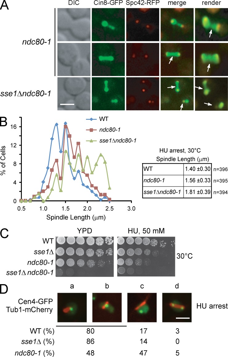
Figure 6.
Cin8-GFP localization pattern in kinetochore mutant ndc80-1 mirrors its localization in the sse1Δ strain. (A) Logarithmically growing cultures of ndc80-1 and sse1Δndc80-1 expressing Cin8-GFP and the SPB marker Spc42-RFP were synchronized in S phase with HU for 2.5 h at 30°C. Images were then obtained using fluorescence confocal microscopy. Arrows point to Cin8 accumulation in the midzone area and the nucleoplasm. DIC, differential interference contrast. Bar, 5 µm. (B) Spindle length in HU-arrested ndc80-1 and sse1Δndc80-1 cells grown at 30°C was measured using Spc42-RFP fluorescence by confocal microscopy. The data shown are from a single representative experiment out of three repeats. (C) 10× serial dilutions of log-phase cells of the indicated genotypes were spotted onto YPD and incubated at 30°C for 2 d. (D) The localization of Cen4-GFP and Tub1-mCherry in HU-arrested WT sse1Δ and ndc80-1 cells is shown. A single GFP dot near one end of the spindle might represent cases in which chromosome IV is attached to one SPB, as not all centromeres are replicated in the presence of HU (a). Cen4-GFP was also observed in the middle of the spindle (b), displaced from the spindle, which might represent detached chromosomes (c), and duplicated on both ends of the spindle (d). The percentages were obtained based on observing 100 cells. Bar, 5 µm.

