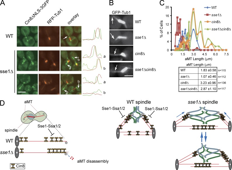
Figure 9.
The effect of Sse1 on Cin8 oligomerization. (A) Representative images of Cin8ΔNLS-3GFP (green) and RFP-Tub1 (red) expressed in WT and sse1Δ cells. Arrows indicate the localization of Cin8ΔNLS-3GFP to aMT plus ends. The curves on the right show the intensity of the GFP and RFP signals along the spindle. Bar, 5 µm. (B and C) Logarithmically growing cultures of WT, sse1Δ, cin8Δ, and sse1Δcin8Δ cells expressing a plasmid-borne copy of GFP-Tub1 were synchronized in S phase with 100 mM HU for 2.5 h at 26°C. aMT length was measured using GFP-Tub1 and/or Spc42-RFP fluorescence by confocal microscopy. Arrows in B point to aMT in the cytoplasm. Bar, 5 µm. The data shown in C are from a single representative experiment out of three repeats. (D) A model for the regulation of Cin8 motility on MT by Sse1-Ssa1/2. Refer to the text for further details.

